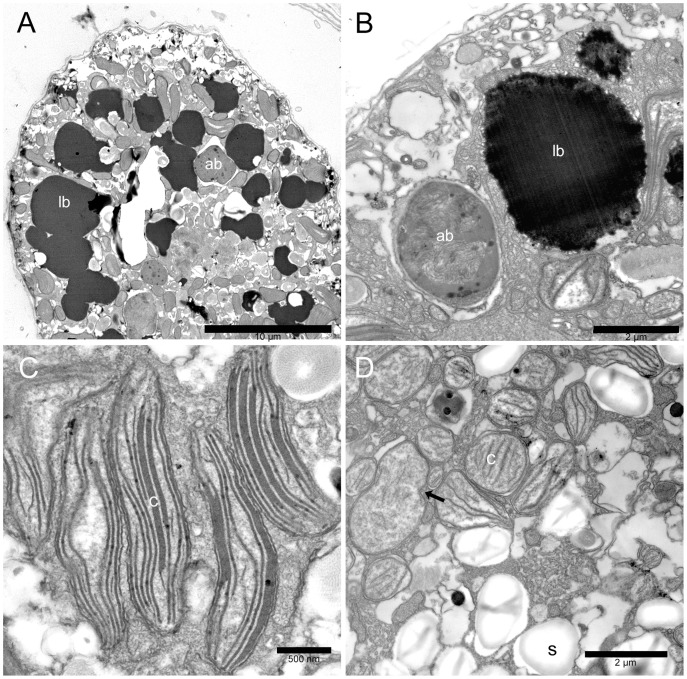
Figure 5. Ostreopsis cf. ovata lipid droplets, accumulation bodies, and chloroplasts: Transmission electron microscopy.
(A) Cell section with numerous large electron dense lipid droplets (lb) in the peripheral cytoplasm. An accumulation body (ab) is visible. Fixation 2. Scale bar 10 µm. (B) Lipid droplet (lb) and accumulation body (ab) in the peripheral cytoplasm. Lipid droplets are highly osmiophilic and present an irregular contour with small drops coalescing together. The accumulation body contains membranous and fibrillar material and appears to be surrounded by smooth endoplasmic reticulum. Fixation 1. Scale bar 2 µm. (C) Elongated chloroplasts (c) in the peripheral cytoplasm. Fixation 1. Scale bar 500 nm. (D) Small rounded plastids (c) with single thylakoids and girdle lamella in the inner part of the cell. A dividing plastid is visible (arrow). Starch granules (s). Fixation 1. Scale bar 2 µm.

