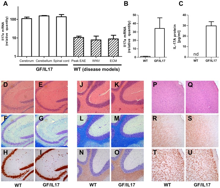
Figure 1. GF/IL17 mice express IL-17A mRNA and protein without major histological defects or leukocyte infiltration.
(A) Relative expression of Il17a mRNA in GF/IL17 mice is equally distributed between forebrain, hindbrain and spinal cord. In comparison to disease models in WT mice (peak EAE, West Nile Virus encephalitis, experimental cerebral Malaria) CNS expression levels of Il17a in otherwise untreated GF/IL17 mice exceed levels of all tested disease models irrespective of the cellular source. (B) Il17a mRNA expression from GF/IL17 primary astrocytes and WT controls was quantified using real time PCR. (C) Astrocyte secretion of IL-17A protein was confirmed using ELISA. Supernatants of GF/IL17 and WT control primary astrocytes were analyzed after 12 hours of culture. In supernatant of WT control astrocytes IL-17A protein was not detectable. (D–U) Routine histological characterization of mice excluded major tissue damage or leukocyte infiltration. Representative areas of hippocampus (D–I) , cerebellum (J–O), or cortex (P–U) are shown in WT controls (D, F, H, J, L, N, P, R, T) and GF/IL17 transgenic mice (E, G, I, K, M, O, Q, S, U) by HE (D, E, J, K, P, Q), LFB (F, G, L, M), anti murine NeuN mAb (H, I, T, U), or anti murine PLP mAb (N, O, R, S) staining (age 9 month).

