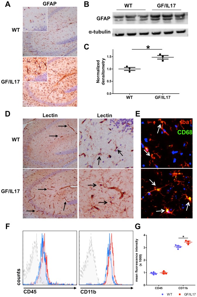
Figure 2. Transgenic CNS expression of IL-17A induces glial activation.
. (A) IHC for GFAP in the hippocampus of WT and GF/IL17 transgenic mice at 9 month. GFAP-staining revealed a strong astrocytic activation by morphological criteria in GF/IL17 mice. (B) Astrocytosis was confirmed by anti-GFAP immunoblotting. Whole brain lysates were analyzed by immunoblotting for the presence of GFAP. Anti-tubulin immunoblotting served as internal loading control on the same membrane. (C) Densitometric quantifications (arbitrary densitometry units) from immunoblots of B after normalization by tubulin densitometry units obtained from the same immunoblot. (*p < 0.05). (D) Tomato-lectin-staining in the hippocampus revealed an activated microglial morphology in GF/IL17A transgenic animals characterized by rounded cell bodies and microglial clustering (open arrows). In addition Lectin staining displayed prominent microvasculature in GF/IL17 mice compared with WT controls (closed arrows; see also Figure 3 for vascular pathology). (E) IHC of frozen brain sections for Iba1 (red), CD68 (green) and Dapi (blue). GF/IL17 mice showed a strong immunoreactivity for the activation marker CD68 in Iba1 stained microglia (white arrows indicating colocalisation of the lysosomal markes CD68 and Iba1, age: 9 month). (F) Representative flow cytometric analysis of surface marker expression from freshly isolated microglia in GF/IL17 mice (red) and WT littermate controls (blue). Dashed histogram: isotype control. Histograms were gated on microglial population according to forward/side scatter profile. GF/IL17 mice displayed similar surface expression levels for CD45 compared with WT whereas CD11b expression was upregulated in GF/IL17 mice compared with WT. (G) Statistical analysis of mean fluorescence intensity of freshly isolated microglia in GF/IL17 mice (red) and WT littermate controls (blue). Comparable expression levels of CD45 in GF/IL17 and WT mice whereas CD11b expression levels were significantly upregulated in GF/IL17 mice compared with WT controls (*p < 0.05).

