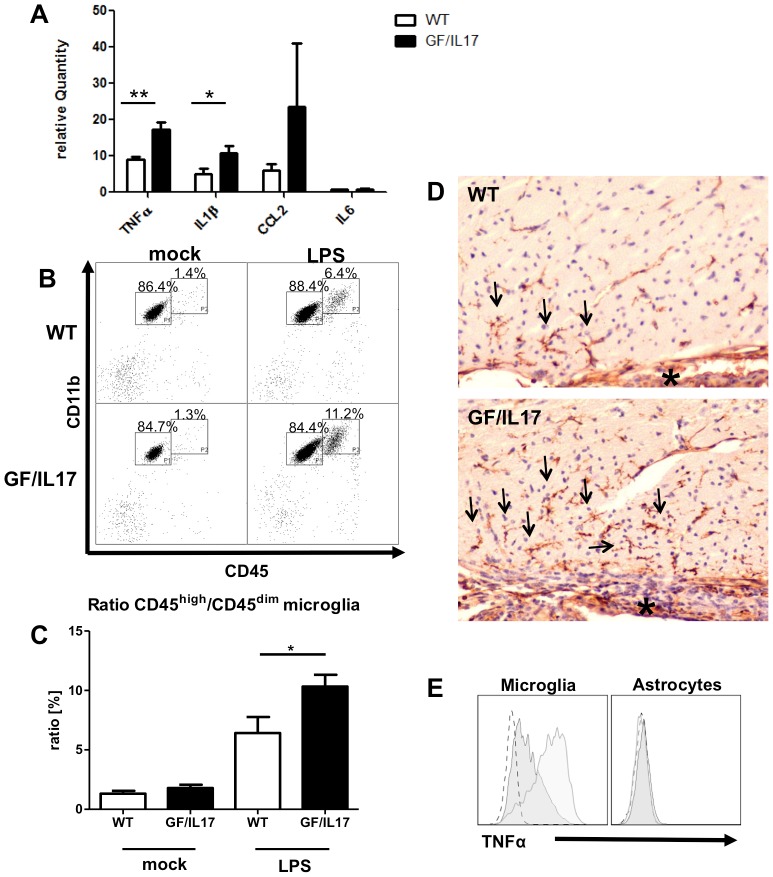
Figure 4. Transgenic IL-17A acts synergistically with other inflammatory stimuli and potentiates LPS induced microgial activation.
GF/IL17 transgenic animals and littermate controls between 2 and 3 month were injected twice with 50 µg LPS i.p. in 24 hours or treated with mock injections of PBS. (A) Quantitative rt-PCR revealed a strong upregulation of the expression of inflammatory cytokines Tnf, Il1b, and Ccl2 by LPS treatment whereas Il6 was not induced following endotoxemia. This effect was markedly pronounced in GF/IL17 transgenic animals (Tnf: p < 0,01; Il1b: p < 0,05). Furthermore Ccl2 expression was strikingly upregulated in some of the LPS treated GF/IL17 mice compared with LPS treated wild-type controls but due to the high interindividual variance not considered as significant. (B) Representative flow cytometry profiles from mock- or LPS-treated GF/IL17 and WT mice. LPS treatment induced a population of CD45high/CD11b+ activated microglia in both WT and GF/IL17 mice. Chronic IL-17A stimulation strikingly augmented this effect, respectively. The numbers above the indicated gate show the mean percentages of FSC/SSC gated populations. (C) For the statistical analysis of infiltrating cell numbers a ratio between CD45high/CD11b+ activated microglia and CD45dim/CD11b+ resting microglia was calculated for each individual mouse.GF/IL17 transgenic animals exhibited a significantly elevated ratio of CD45high/CD11b+ activated to CD45dim/CD11b+ microglia after LPS treatment, respectively (p < 0,05). (D) Lectin immunohistochemistry revealed a pronounced accumulation of activated microglia (arrows) in the periventricular regions (asterisk: choroid plexus) after LPS treatment in GF/IL17 mice. (E) Intracellular staining of TNF-α after LPS treatment. Only CD45 positive microglia and monocytes/macrophages are stained by anti TNF-α antibody (left histogram) whereas GLAST positive astrocytes are negative for TNF-α (right histogram). In GF/IL17 transgenic mice (light gray) compared with wildtype mice (dark gray) CD45 positive microglia and monocytes/macrophages exhibit a stronger intracellular TNF-α staining after LPS treatment.

