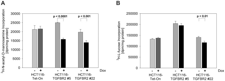
Figure 4. Incorporation of 3H-labeled monosaccharides.
Radioactive labeling experiments were performed in the presence and absence of dox (0.5 µg/ml) and by exposure to TGF-ß1 (10 ng/ml) for 72h. (A) Incubation with 3H-ManNAc resulted in a significant reduction of incorporated ManNAc in the TGFBR2 clones #5 and #22 but not in the parental HCT116-Tet-On cell line. (B) Incorporation of 3H-L-fucose was slightly reduced in presence of dox in both TGFBR2 clones in contrast to HCT116-Tet-On cells. Values represent the mean of three independent experiments ±S.D.

