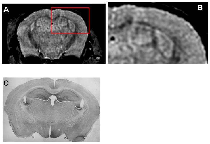
Figure 4. Shows an in vivo μMRI of a 16 month old wild-type mouse after intravenous femoral injection of USPIO-PEG-Aβ1-42.
B shows a higher magnification of the area in the red box of shown in A. Some dark spots are also evident in A (likely corresponding to blood vessels) but far fewer than seen in Figure 3A or Figure 4A . C shows the matching tissue section to A immunolabeled with anti-Aβ 4G8/6E10. No amyloid deposits are evident, as expected.

