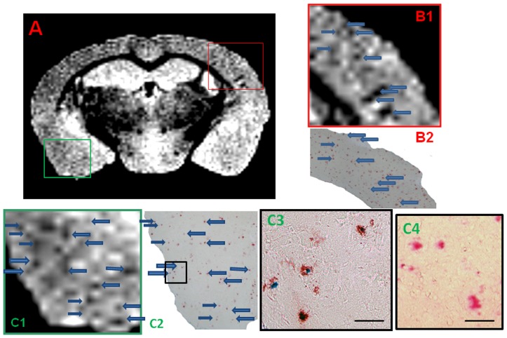
Figure 5. Shows an ex vivo μMRI coronal cut at the level of the anterior hippocampus of a 15 month-old APP/PS1 Tg injected with USPIO-PEG-Aβ1-42, showing numerous dark spots.
In B1 a higher magnification of the μMRI in the red box in A is shown. This is matched to amyloid plaque immunolabeling (in red) shown in B2. Arrows highlight some matches between the μMRI and histology. Figure C1 is higher magnification of the μMRI in the green box in A. This is matched to double amyloid plaque immunolabeling and Perl iron staining in C2 and C3. Arrows highlight some of the matches between the μMRI in C1 and immunolabeling in C2. C3 is a higher magnification of the black box in C2. C4 shows and example of double amyloid plaque immunolabeling and Perl iron staining in an APP/PS1 mouse injected with USPIO alone. Only the red immunolabeling of amyloid plaques is seen with no co-label of iron. Co-labeling of amyloid plaques with anti-Aβ 4G8/6E10 (in red) and Perl iron stain (in blue) highlights the co-deposition of USPIO-PEG-Aβ1-42 on amyloid deposits in C2 and C3 (scale bar = 100 microns).

