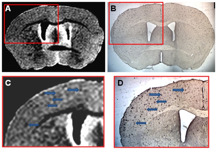
Figure 6. Shows an example of ex vivo μMRI, at the level of the area cinguli of the cerebral cortex, of a 14 month-old APP/PS1 Tg injected with USPIO-PEG-Aβ1-42, showing numerous dark spots.
This is matched to a tissue section immunolabeled with anti-Aβ 4G8/6E10 in B. C is higher magnification of the μMRI in the red box in A, while D is a higher magnification of the tissue section immunolabeling seen in B.

