
Fig. 2.
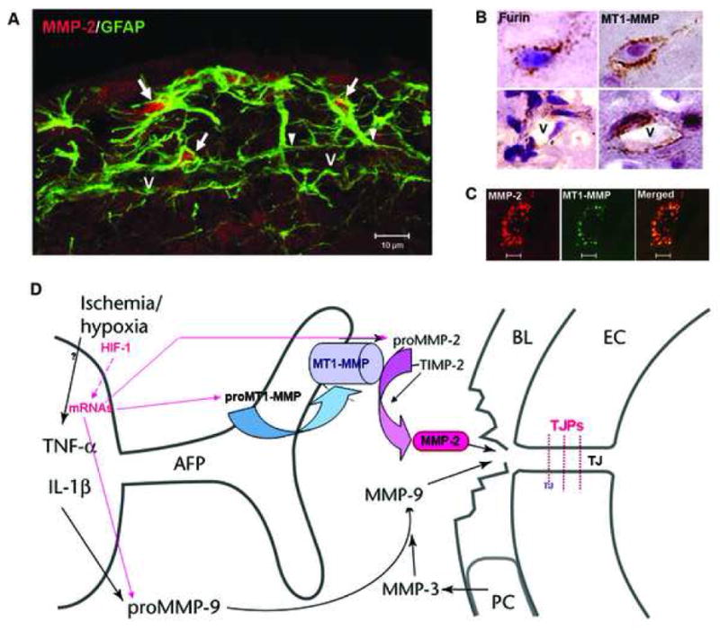
(A) Confocal immunohistochemistry shows GFAP-positive astrocytes around a vessel (V) that express MMP-2 (arrows) in intact rat brain tissue. The arrowheads indicate the astrocyte endfeet around the vessel. (B) Expression of furin and MT1-MMP in brain cells and around vessels (V). (C) Confocal images show the co-localization of MMP-2 and MT1-MMP immunohistochemistry in brain cells. (D) Schematic drawing to show that the activation of MMP-2 occurs through the action of the trimolecular complex during the early opening of the BBB in 3h-reperfusion after 90 min MCAO. In the astrocytic foot processes (AFP), the membrane-type 1 matrix metalloproteinase (MT1-MMP) joins with tissue inhibitor of metalloproteinases-2 (TIMP-2) to activate proMMP-2 in a spatially constrained manner close to the basal lamina (BL). In the BL are the pericytes (PC). The endothelial cells (EC) have tight junctions (TJ). The activated MMP-2 has direct access to the portion of the BL beneath the AFP and components of the BL are degraded. The manner in which this disruption of the BL leads to increased permeability is unclear since the role of the BL in maintaining the integrity of the blood vessel is uncertain.