
Fig. 4.
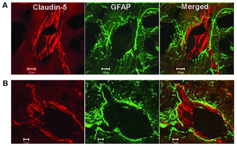
Confocal micrographs showing claudin-5 immunoreactivity after 3 h of reperfusion following 90 min of MCAO in the rat. (A) The nonischemic side show that the claudin-5 (Cy-3) in blood vessels is separated from the astrocytes (GFAP-FITC) surrounding them. The merged images show that the claudin-5 and astrocytes are separate. (B) In the ischemic hemisphere, there is fragmentation and degeneration of the claudin-5 immunoreactivity. Co-localization of claudin-5 and GFAP was seen in the ischemic hemisphere. Adapted from Yang et al., 2007.