
Fig. 7.
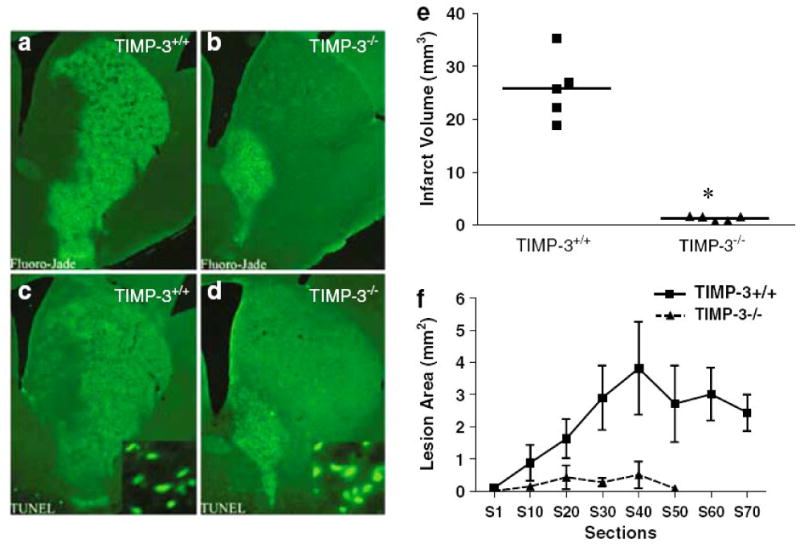
Lesion size in timp-3 +/+ versus timp-3 −/− at 3 days of reperfusion following 30 min transient MCAO. Timp-3 +/+ and timp-3 −/− mice were subjected to 30 min transient MCAO followed by 3 days of reperfusion. Coronal sections were stained for degenerating neurons using Fluoro-Jade (a, b) or stained for apoptotic nuclei using TUNEL (c, d). Infarct volume in timp-3 +/+ versus timp-3 −/− mice as assessed by morphometric analysis of Fluoro-Jade-stained histological sections, (e) n=5 mice per strain, *P<0.001. (f) Average lesion area per histological section in rostrocaudal direction in timp-3 +/+ versus timp-3 −/− mice. Taken from Wetzel et al., 2008.