
Fig. 1.
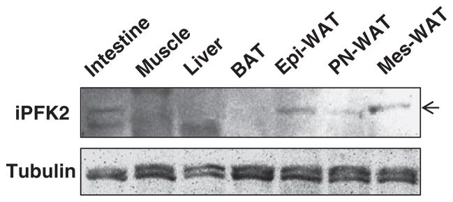
PFKFB3/iPFK2 expression in key metabolic tissues. Male wild-type C57BL/6J mice were fed ad libitum. At 12–14 weeks of age, mice were euthanized for collection of tissue samples. Tissue lysates were prepared to determine the amount of iPFK2 using Western blot analyses. BAT, brown adipose tissue; Epi-WAT, epididymal white adipose tissue; PN, perinephric; Mes, mesenteric.