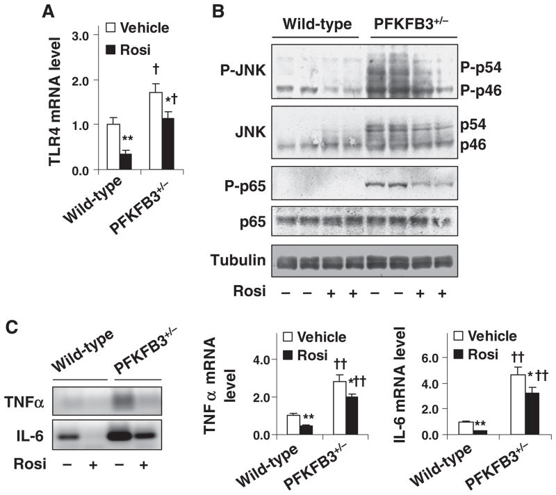
Fig. 4.
Involvement of PFKFB3/iPFK2 in the effect of PPARγ activation on diet-induced intestine inflammatory response. Male PFKFB3+/− mice and wild-type littermates, at 5–6 weeks of age, were fed an HFD for 12 weeks and treated with rosiglitazone (Rosi, 10 mg/kg/day in PBS) or vehicle (PBS) orally for the last 4 weeks of the feeding regimen. (A) Intestine mRNA levels of TLR4 were quantified using real-time RT-PCR. (B) Intestine inflammatory signaling. Intestine lysates were prepared to determine the amount and phosphorylation states of JNK and NF-κB p65 using Western blot analyses. (C) Intestine mRNA levels of TNFα and IL-6 were quantified using real-time RT-PCR. Left panel, representative PCR products; and right two panels, relative intestine mRNA levels. For panels (A) and (C), numeric data are means±S.E.; n=4–6. *P<.05 and **P<.01, rosiglitazone vs. vehicle for the same genotype; †P<.05 and ††P<.01, PFKFB3+/− vs. wild type for the same treatment (rosiglitazone or vehicle).

