
Figure 4.
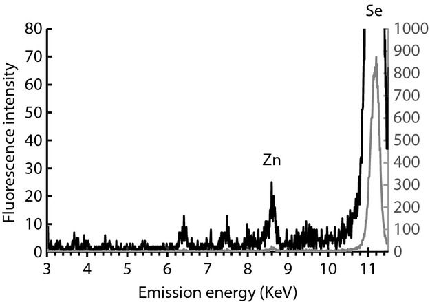
X-ray fluorescence scan of Se-Met-labeled PilD. Sample was prepared as described in the Experimental Procedures, with selenomethionine in place of methionine in the amino acid mix. In this X-ray fluorescence scan for elemental analysis, excitation at 12,660 eV allowed observation of the strong Se emission peak from the 12 methionines in PilD as well as the Zn emission peak. Identical data are plotted on two scales to highlight the Zn signal.