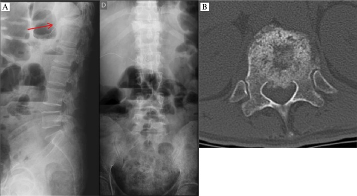
Figure 1.
Example of a clinical case used in the evaluations. “Forty one-years-old patient with a diagnosis of metastatic colon adenocarcinoma. He has a complaint of progressive dorsal pain which is worse at night and with movement. The patient has a limited ability to move on the bed due to dorsal pain.” A. Anteroposterior and profile radiographs. B. Axial cut in computed tomography showing the lesion site.

