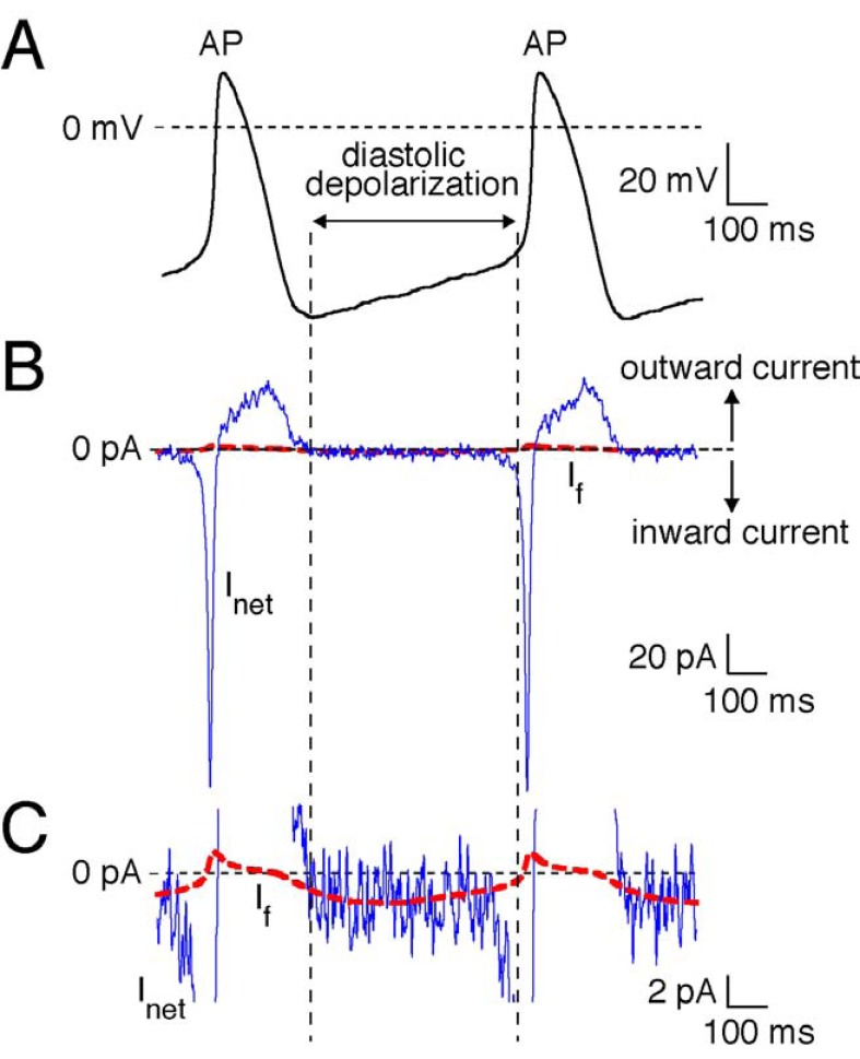
Fig. (2).
Experimentally recorded action potential (AP) of a single human SAN pacemaker cells.
A. For cell isolation and recording procedure, see Ref 25. B. Associated net membrane current (Inet, blue line) calculated from Inet=- Cm×dVm/dt, where Cm and Vm denote membrane capacitance and membrane potential, respectively. Note the small inward current underlying the slow diastolic depolarization. C. Computed If (solid red line). The time course of If was reconstructed using the Vm values of the recorded action potentials and a first-order Hodgkin–Huxley type kinetic scheme based on voltage clamp data from human SAN pacemaker cells. For details, see Ref 24.

