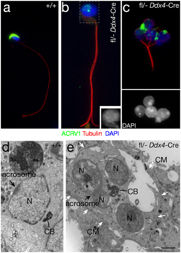
Fig. 2.
Structural analysis of multi-nucleated germ cells from the testes of 2-month-old Myh10fl/− Ddx4-Cre mice. (a–c) Immunofluorescence analysis of testicular germ cells, stained with an antibody against ACRV1 (green), a component of the acrosome (Reddi et al., 1995), and with an anti-tubulin antibody to visualize the axoneme (red). Nuclei are stained with DAPI (blue). (a) Wild type sperm. (b) MYH10-deficient germ cell with a bundle of four flagella. Inset: grayscale image showing the head (boxed area) with DAPI stained nuclei and four flagella (four bundles of microtubules). (c) MYH10-deficient germ cell with four nuclei (DAPI staining shown in bottom panel), bundles of microtubules, and defective acrosomes. (d and e) Ultrastructural analysis of a wild type spermatid (d), and a MYH10-deficient germ cell with four nuclei (e). N, nucleus; CB, chromatoid body; CM, cell membrane. White arrows mark the cell membrane. Black arrow marks the acrosome. Scale bar (d and e), 2 µm.

