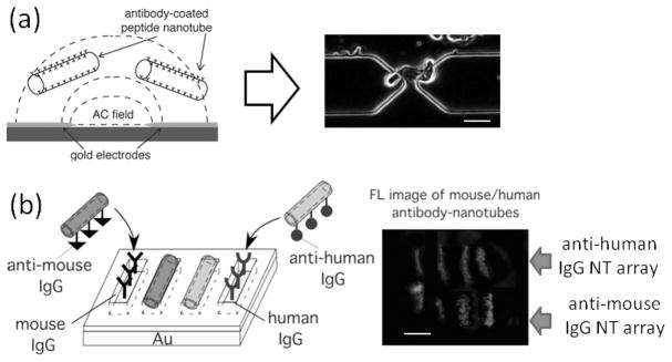
Figure 2.

(a) Peptide nanotubes are injected onto the electrode-patterned platform while applying an AC field (left), and then peptide nanotubes are trapped at the gap between adjacent electrodes by positive dielectrophoresis (right in an optical micrograph). Scale bar = 2 μm. (b) (left) Scheme to assemble anti-mouse IgG-coated nanotubes and anti-human IgG-coated nanotubes onto their antigen-patterned substrates via biomolecular recognition; Location-specific immobilization of Alexa Fluor 546-labeled anti-mouse IgG nanotubes onto the mouse IgG trenches and FITC-labeled anti-human IgG nanotubes onto the human IgG trenches. (right) Fluorescence image of anti-mouse IgG nanotubes (in red) and anti-human IgG nanotubes (in green), attached onto four upper trenches filled with mouse IgG and four bottom trenches filled with human IgG, respectively, scale bar = 2 μm. (a, reproduced with permission from ref. 23, copyright Wiley-VCH Verlag GmbH & Co. KGaA. b, reprinted with permission from ref. 10, copyright 2005 American Chemical Society)
