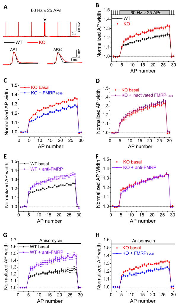
Figure 1. FMRP Regulates Action Potential Duration in CA3 Pyramidal Neurons in a Translation-Independent and Cell-Autonomous Presynaptic Manner.
(A) Sample traces of AP trains recorded in CA3 PCs of WT (in black, here and throughout) or Fmr1 KO mice (in red, here and throughout) in response to 25 stimuli at 60 Hz. 4 APs at 0.2 Hz were evoked prior to each train to determine baseline AP duration. Blue lines show the AP width measured at −10 mV level.
(B) Analysis of AP duration in (A). AP duration was normalized to its own baseline for WT or Fmr1 KO mice.
(C) Effect of intracellular perfusion of FMRP1-298 in Fmr1 KO CA3 PCs on AP broadening.
(D) Heat-inactivated FMRP1-298 failed to affect AP broadening in Fmr1 KO mice.
(E) Effect of intracellular perfusion of anti-FMRP Ab in WT CA3 on AP broadening.
(F) Intracellular perfusion of Anti-FMRP Ab had no effect on AP duration in Fmr1 KO mice.
(G, H) Anisomycin failed to block the effects of anti-FMRP on AP duration in WT mice (G), or the effects of FMRP1-298 on AP duration in Fmr1 KO mice (H).

