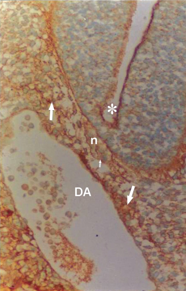
Fig 2.

Photomicrograph of GD10 mouse embryo, cross-section of paraxial mesenchyme, notochord, and ventral portion of neural tube incubated with WFA lectin. Notochordal plate (n), its sheath (small arrow), neural tube (star), DA; Dorsal aorta. Severe reaction observed in paraxial mesenchyme (arrow; ×200).
