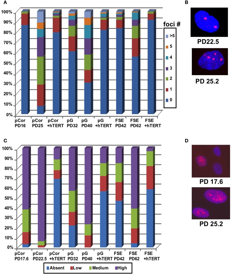
Figure 4.
ICF fibroblasts display high levels of γ-H2AX foci and p21 staining at abnormally low population doublings. (A) pCor, pG and FSE fibroblasts with and without expression of hTERT were stained with an antibody to γ-H2AX. Cells were scored for the number of nuclear foci. (B) Representative pCor nuclei at PDs 22.5 and 25.2 illustrate γ-H2AX foci. (C) pCor, pG, and FSE fibroblasts with and without expression of hTERT were stained with an antibody for p21 and were scored for staining intensity. (D) Representative pCor nuclei at PD 17.6 and 25.2 demonstrating p21 staining.

