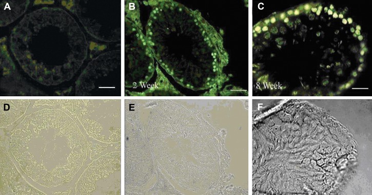
Fig 1.
Donor-derived spermatogonial cells were traced in the recipient testes: A. The control group without addition of primary antibody; B, C. Donor-derived spermatogonial cells were traced in the recipient testes, two and eight weeks after transplantation, respectively. The cells showing nuclear BrdU staining were considered as transplanted cells. D, E and F consist of phase-contrast photographs. Magnification ×400 (in A, B); ×800 (in C) (bar =10 µm)

