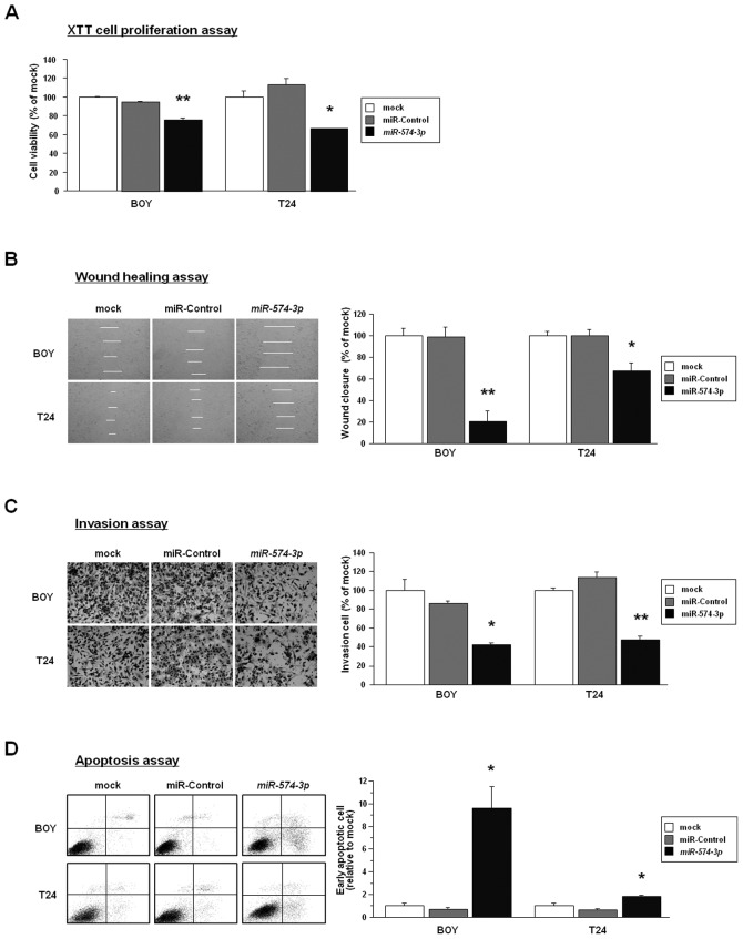
Figure 2.
Gain-of-function studies in miR-574-3p-transfected BC cell lines. (A), Cell proliferation determined by the XTT assays of the transfectants. *P<0.001. **P<0.0001. (B), Wound healing assays demonstrated significant inhibition of cell migration in miR-574-3p transfectants. Phase contrast micrographs of the transfectants (BOY and T24) taken at 0 and 24 h after monolayer wounding are shown on the left. Quantification of cell migration using the monolayer wound healing assay is shown on the right. *P<0.001. **P<0.0001. (C), Significant inhibition of cell invasion was observed in miR-574-3p transfectants. Phase contrast micrographs of invading transfectants (BOY and T24) are shown on the left. Quantitation of cell invasion is shown in the right panel. *P<0.01. **P<0.0001. (D), Apoptosis assay by flow cytometry. Significant numbers of early apoptotic cells were observed in miR-574-3p transfectants. Early apoptotic cells can be seen in the bottom right quadrant. The percentages of early apoptotic cells in the miR-control and the miR-574-3p transfectants are shown in the histogram. *P<0.01.

