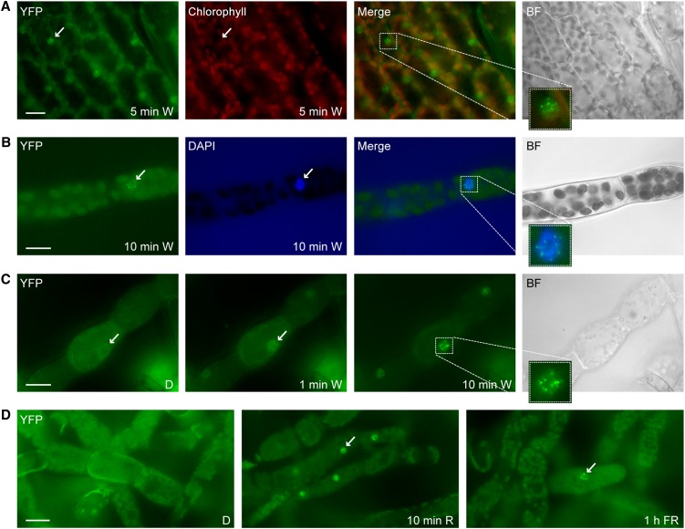
Figure 1.
Rapid Light-Induced Nuclear Transport of P. patens PHY1.
(A) Light-regulated nuclear accumulation of Pp-PHY1 in gametophores. Dark-adapted gametophores of transgenic P. patens plants expressing Pp-PHY1:YFP were exposed to W light for 5 min and used for fluorescence microscopy.
(B) DAPI staining. Dark-adapted protonema filaments of Pp-PHY1:YFP expressing P. patens plants were exposed to W light for 10 min, fixed with formaldehyde, stained with DAPI, and analyzed by fluorescence microscopy.
(C) Rapid light-induced nuclear transport of Pp-PHY1 in protonema filaments. Dark-adapted protonema filaments of P. patens plants expressing YFP-tagged Pp-PHY1 were used for time series fluorescence microscopy. Images were acquired before (dark control [D]) and 1 and 10 min after the onset of irradiation with W light.
(D) Nuclear accumulation of P. patens PHY1 is induced by R and FR light. Protonema filaments of P. patens plants expressing Pp-PHY1:YFP were dark adapted and used for fluorescence microscopy. Images were acquired before and after irradiation with R light (10 min, 22 μmol m−2 s−1) or FR light (1 h, 18 μmol m−2 s−1). The samples were fixed with formaldehyde before microscopy analysis.
Arrows indicate nuclei; insets show enlargements of nuclei. Merge, merge of YFP and chlorophyll/DAPI channels; BF, bright field. Bars = 20 μm.

