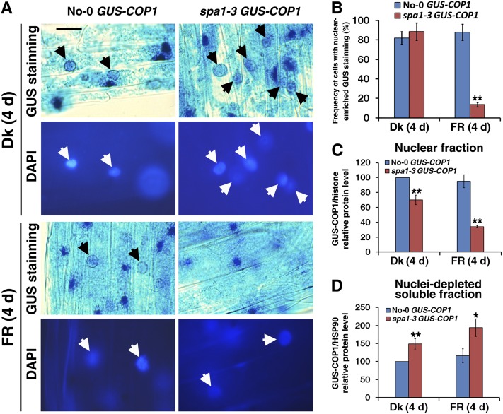
Figure 8.
Nuclear Accumulation of COP1 Is Defective in the spa1-3 Mutant under FR Light.
Seedlings were grown in the dark (Dk) or FR light (2.5 µmol·m–2·s–1) for 4 d.
(A) Nuclear accumulation of GUS-COP1 in the parental No-0 GUS-COP1 plants (left panels, indicated by black arrows) but not in the spa1-3 GUS-COP1 plants (right panels) under FR light. The top panels present GUS-stained images and the bottom panels display DAPI staining of the corresponding image to show the positions of the nuclei (white arrows). The unmarked blue GUS staining dots are GUS-COP1 cytoplasmic inclusion bodies. Bar = 20 μm.
(B) Difference in frequency of cells with nuclear-enriched GUS staining in No-0 GUS-COP1 and spa1-3 GUS-COP1 in the dark or under FR light.
(C) Quantification of relative GUS-COP1/histone protein levels in the purified nuclear fraction corresponding to Supplemental Figure 6C online, indicating reduced nuclear abundance of GUS-COP1 in the spa1-3 mutant background. Error bars represent sd from triplicate experiments. Asterisks mark significant differences in relative GUS-COP1/histone protein levels from No-0 GUS-COP1 according to Student’s t test (*P < 0.05; **P < 0.01).
(D) Quantification of relative GUS-COP1/HSP90 protein levels in the nuclei-depleted soluble fraction in response to FR light, corresponding to Supplemental Figure 6D online. Error bars represent sd from triplicate experiments. Student’s t tests were performed as in (C) (see Supplemental Figure 6 online).

