Figure 4.
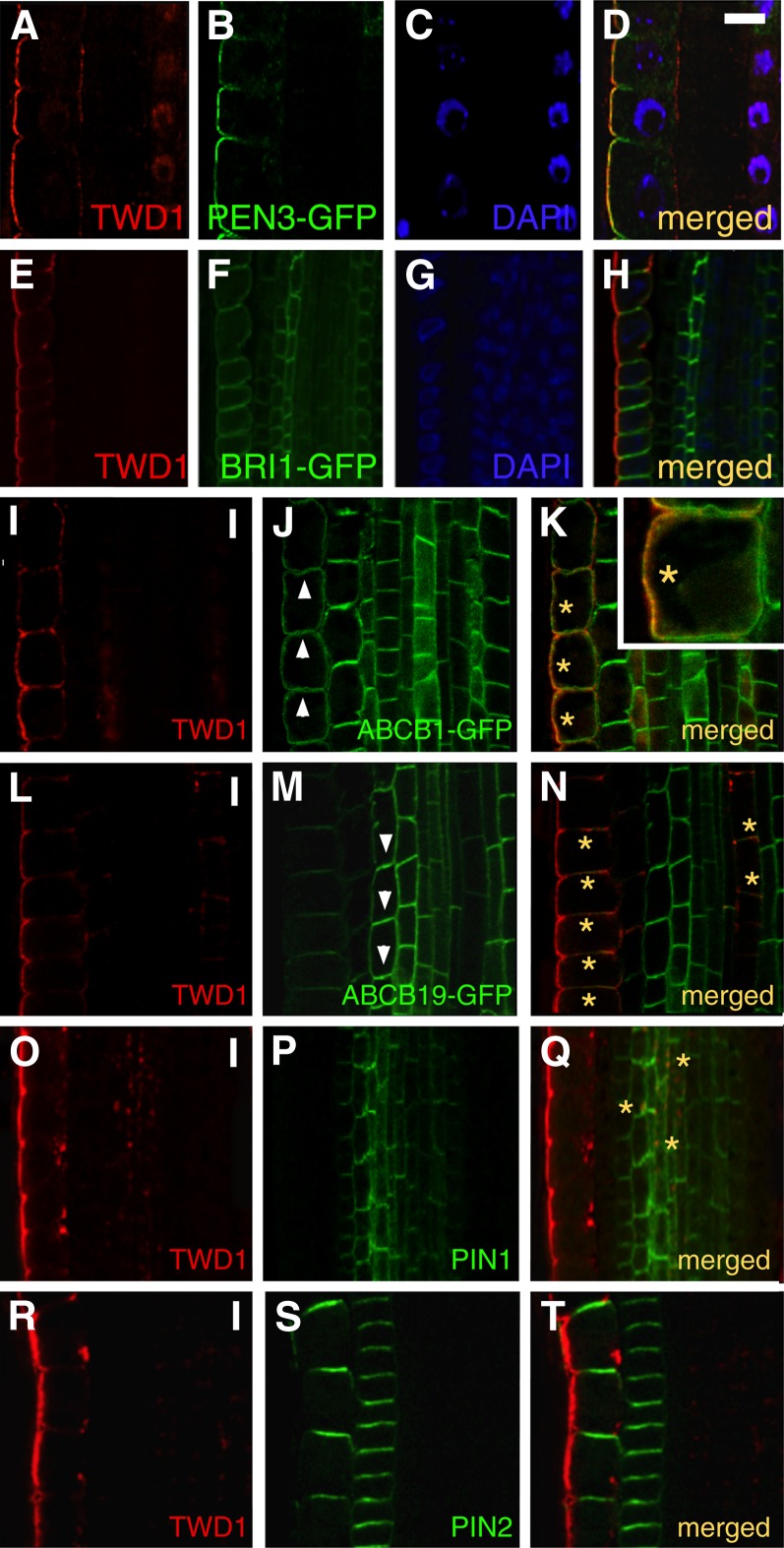
TWD1 Reveals a Lateral PM Location in the Root Epidermis Where It Colocalizes with PEN3/ABCG36 and ABCB1.
(A) to (H) Whole-mount immunostaining using anti-TWD1 with PM markers PEN3/ABCG36-GFP ([A] to [D]) and BRI1-GFP ([E] to [H]). Note predominant lateral PM localization ([A]; stronger at the outer polar domain) in the root epidermis, which matches outward-facing, lateral domains of polar marker PEN3/ABCG36-GFP (D) but only partially overlaps the nonpolar marker BRI1-GFP (H). Note that a weak nuclear TWD1 signal in (A) is probably caused by bleed-through of the DAPI signal because it was not found without DAPI. Bar = 15 μm.
(I) to (T) TWD1 colocalizes with ABCB1-GFP ([I] to [K]) and occasionally also with ABCB19-GFP ([L] to [N]) on lateral and basal domains in the epidermis and stele, respectively. Note only partial overlap with PIN1-GFP ([O] to [Q]) and absence of overlap with PIN2-GFP ([R] to [T]). Arrowheads indicate directions of main shootward (J) and rootward (M) auxin flows in the epidermis and endodermis, correlating with main B1 (J) and B19 (M) expression, respectively. Note that B1-TWD1 and B19-TWD1 colocalization is limited to subdomains in the epidermis ([K] and [N]) and stele (N), respectively (marked by asterisks). Bars = 10 μm.

