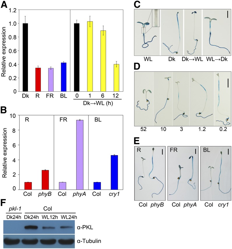
Figure 2.
PKL Expression in the Hypocotyls Is Repressed by Light.
(A) A qRT-PCR assay showing PKL transcript levels in wild-type seedlings subjected to red (R), far-red (FR), or blue (BL) light or darkness (Dk) for 5 d or in 5-d-old dark-grown seedlings transferred to white light (WL) for the indicated periods of time (0 to 12 h). Data represent the mean ± sd of three biological replicates.
(B) qRT-PCR assay showing increased PKL expression, relative to the Col wild type, in phyB, phyA, and cry1 photoreceptor mutants grown in red, far-red, and blue light conditions, respectively, for 5 d. For (A) and (B), relative expression was normalized to that of UBQ1, and data represent the mean ± sd of three biological replicates.
(C) GUS staining of ProPKL:GUS transgenic seedlings. Seedlings were grown in white light or darkness for 5 d, 4-d-old dark-grown seedlings were exposed to white light for an additional 1 d (Dk→WL), or 4-d-old light-grown seedlings were transferred to darkness for an additional 1 d (WL→Dk). The inset in the WL panel is an enlargement of the hypocotyl. Bar = 2 mm.
(D) GUS staining gradually increased in the hypocotyls of seedlings grown in a decreasing series of light intensities (μmol m−2 s−1; indicated below) for 5 d. Bar = 2 mm.
(E) GUS staining in the hypocotyls was significantly enhanced in the photoreceptor mutants compared with the Col wild type. Seedlings harboring the ProPKL:GUS reporter were grown under the indicated light conditions for 5 d. Bar = 2 mm.
(F) Immunoblotting of PKL protein. Seedlings were grown in long-day conditions (16 h day/8 h night) for 3 d and were then transferred to darkness or white light for the indicated period of time at the end of the day. Hypocotyls and cotyledons were detached and harvested for protein isolation. Immunoblotting against the tubulin antibody served as a loading control.

