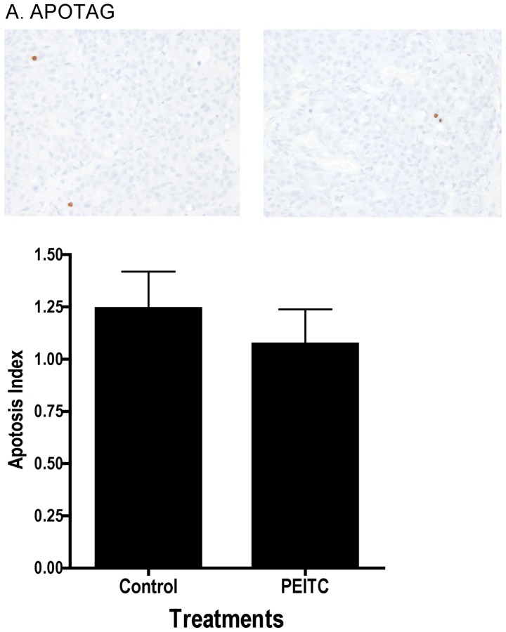
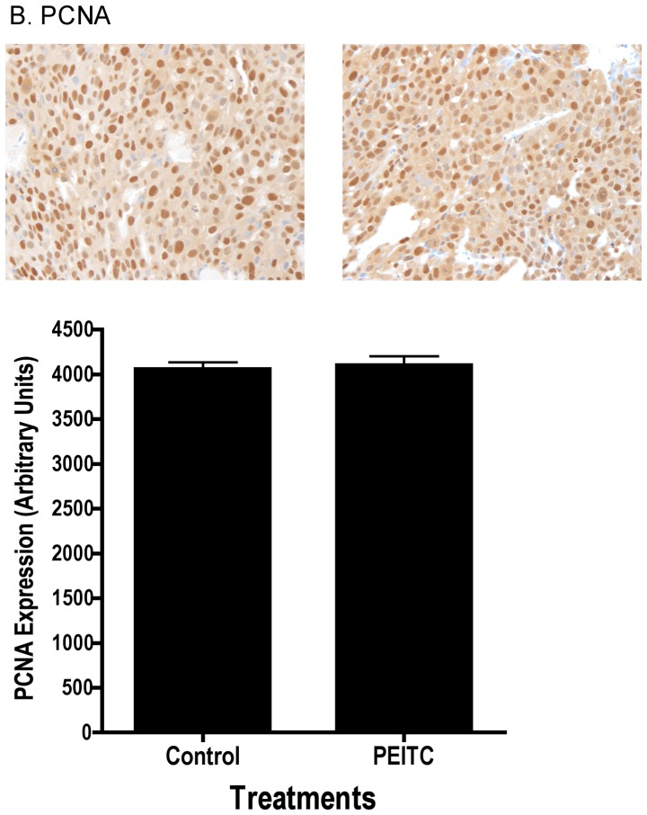
Figure 3.
Dietary administration of PEITC does not induce apoptosis or decrease cellular proliferation in LNCaP prostate cancer cell xenografts in athymic nude mice. Immunohistochemical analysis of markers for apoptosis and proliferation was performed in paraffin-embedded tumor samples from control and PEITC-treated mice. All images were calibrated to a master (single image) color profile to maximize accuracy. (A) ApoTag, apoptosis index = average apoptosis-positive cells/field ± SE. (B) PCNA, proliferation index = average PCNA-positive cells/mm2 ± SE. Representative images of immunohistochmical staining are illustrated on top of bar graphs. Control, n=19; PEITC, n=22.


