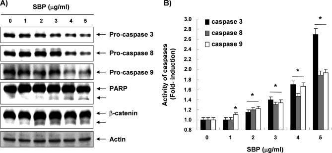
Figure 4.
Activation of caspases and degradation of PARP and β-catenin proteins by SVB treatment in AGS cells. (A) Cells treated with various concentrations of SVB for 24 were lysed and cellular proteins were separated by SDS-polyacrylamide gels and transferred onto nitrocellulose membranes. Membranes were probed with anti-caspase-3, -8, and -9, anti-PARP, and anti-β-catenin antibodies. Proteins were visualized using the ECL detection system. Actin was used as an internal control. (B) Cells grown under the same conditions as (A) were collected and lysed. Aliquots were incubated individually with DEVD-pNA, IETD-pNA, and LEHD-pNA for caspase-3, -8, and -9 at 37°C for 1 h. Released fluorescent products were measured. Data represent the mean of three independent experiments. The statistical significance of results was analyzed by a Student’s t-test (*p<0.05).

