
Figure 1.
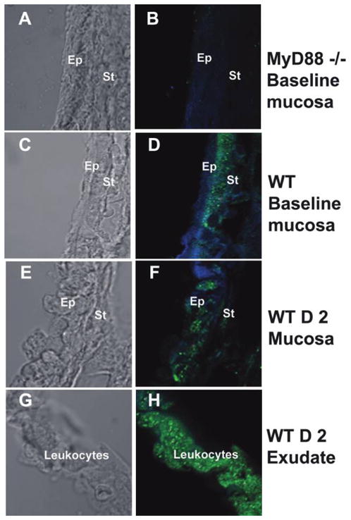
Expression of myeloid differentiation primary response gene 88 (MyD88) in the middle ear (ME). The left column shows light microscopy, and the right column shows immunohistochemical staining with fluorescence. Blue represents cytokeratin, and green represents MyD88. At baseline, MyD88 was not detected in the ME mucosa of MyD88−/− mice (A and B), as at any time point after inoculation with nontypeable Haemophilus influenzae (NTHi). In C57BL/6mice, MyD88 was detected in the stromal layer at baseline (C and D). The boundary between the epithelium and stromal layers is blurred as leukocytes traverse the ME mucosa (E and F). MyD88 was detected throughout the ME mucosa within 2 days after NTHi inoculation in wild-type (WT) mice. MyD88 was strongly detected in leukocytes (identified from adjacent hematoxylineosin–stained sections [data not shown]) comprising the ME exudate (G and H). D 2, day 2; Ep, epithelium; St, stroma.