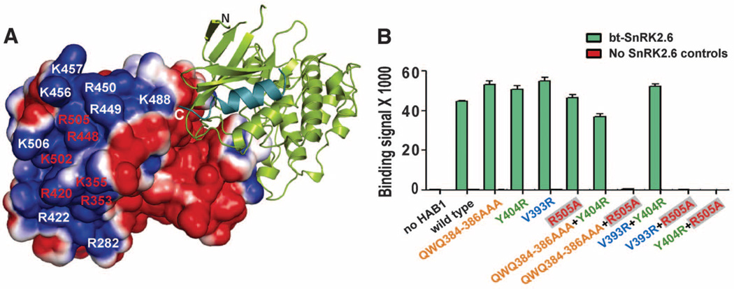
Fig. 3.
SnRK2.6 and HAB1 associate with each other via two modular interactions. (A) Charge-distribution surface of HAB1 in the HAB1-SnRK2.6 complex with an electrostatic scale from –1 to +1 ev, corresponding to red and blue colors. The positively charged residues required for ABA box interaction are labeled in red. (B) Mutational analysis of the HAB1-SnRK2.6 interaction by AlphaScreen luminescence proximity assay. Error bars indicate SD (n = 3). HAB1 mutant proteins are color-coded on the basis of the mutated regions that bind to SnRK2.6. Q384A/W385A/Q386A, which is the “lock” that inserts into both SnRK2.6 and PYL2 ABA receptor, is colored in orange; V393R, which interacts with SnRK2.6 activation loop interaction, is in blue; Y404R, which binds to αG, is in green; R505A, which is required for ABA box interaction, is in red within gray boxes.

