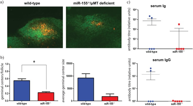
Fig 5.
(a) Immunohistochemistry was performed on mesenteric lymph node sections from wild-type and miR-155/μMT-deficient chimeric mice to detect germinal centers (B220+ cells, green; and PNA+, orange) on day 14 postinfection (p.i.); magnification, ×20. (b) Number of GCs (±SEM) was determined from stained sections. * indicates P value of <0.05 by a Student t test, n = 3 mice per group. Average germinal center size (±SEM) calculated from stained sections in panel b, n = 3 mice per group. (c) Anti-EspA-specific antibody titers were measured in the serum of infected mice 21 days p.i.

