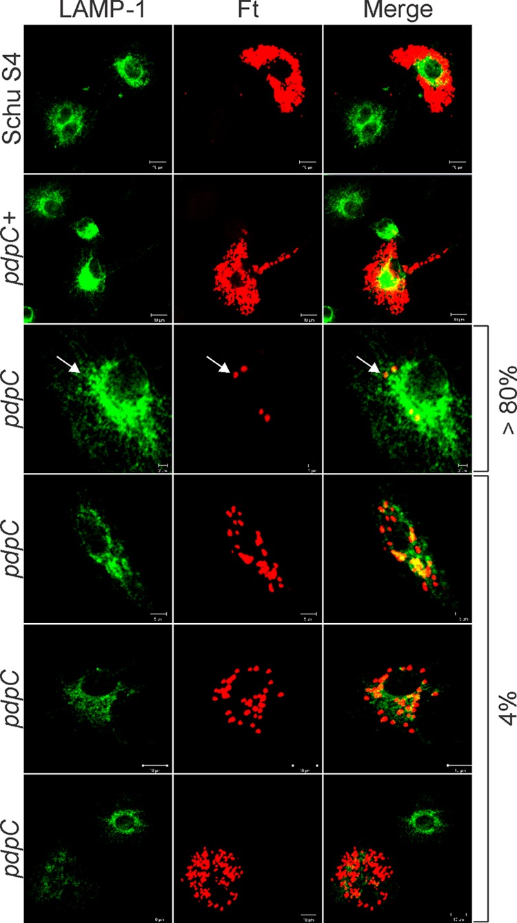
Fig 4.

Two distinct intracellular phenotypes of the pdpC mutant. Shown are confocal images of MDMs infected for 20 h with Schu S4, the pdpC mutant, or the complemented pdpC+ strain. Bacteria are shown in red, and LAMP-1 is shown in green as an indicator of overall macrophage morphology. Schu S4 and the pdpC+ strain grew well in MDMs, whereas the pdpC mutant was defective for growth in 80% of infected cells and grew to a limited extent in ∼4% of infected MDMs, as indicated. The arrows indicate nonreplicating pdpC mutants that do not colocalize with LAMP-1. The data shown are representative of results from more than three independent experiments.
