Fig 2.
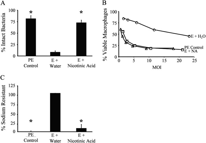
Nicotinic acid treatment induces WT L. pneumophila to express multiple transmissive-phase traits. (A) The extent of lysosome evasion after a 2-h infection of macrophages with L. pneumophila at an MOI of 1 was quantified as the percentage of intracellular bacteria that were intact, observed by fluorescence microscopy. Displayed are the mean results from duplicate samples in three independent experiments. Error bars indicate standard deviations (SD), and asterisks designate significant differences (P < 0.01) compared to the results for the E-phase water control. (B) Macrophage viability after 1-h infection of macrophages at the multiplicities of infection (MOI) shown was quantified with PE-phase (triangles) or E-phase cultures supplemented with water (H2O) or nicotinic acid (NA) using the colorimetric dye alamarBlue. The values plotted represent the means ± SD for triplicate samples determined in one of three similar experiments. At similar MOIs, E-phase cultures supplemented with NA were significantly different than E-phase cultures supplemented with water (P < 0.05). E-phase cultures treated with NA were not statistically different than PE-phase bacteria (P > 0.05). (C) The percentage of sodium-resistant bacteria was measured by plating cultures on medium with or without 100 mM NaCl and then calculating CFU as described previously (41). Values shown = mean [(E + NA or PE control)/(E + H2O) × 100]. Error bars represent SD from duplicate samples in three independent experiments; for E-phase + water results, the error bar is too small to be visible. Asterisks denote statistically significant differences (P < 0.01) compared to the results for the E-phase water control.

