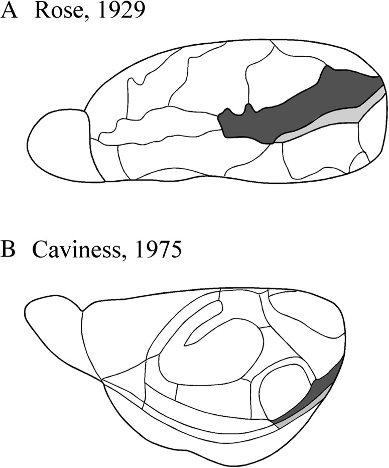
Figure 1.
Historical views of the mouse cortical mantle. (A) Lateral surface view of the mouse brain adapted from Rose (1929). Ectorhinal cortex is shown in dark gray and perirhinal cortex is shown in light gray. (B) Dorsolateral surface view adapted from Caviness (1975). Area 36 is shown in dark gray and area 35 is shown in light gray. Note that the perspectives differ in these 2 views. Both views describe the rostral limit of the perirhinal areas as arising at the caudal limit of the claustrum. The dorsal border is more dorsal in Rose (1929), though this is overemphasized, here, because of differences in perspective. See text for details about nomenclature.

