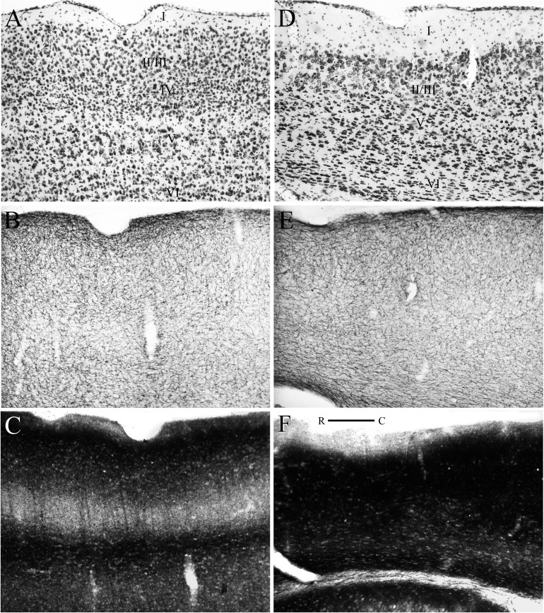
Figure 6.
Horizontal sections showing the cytoarchitecture and histochemistry of perirhinal areas 36 (left panels) and 35 (right panels). Adjacent sections were stained for Nissl material (A and D), the enzyme acetycholinesterase (B and E), and heavy metals using the Timm’s method (C and F). Note the vestigial granular layer in area 36, panel A. Abbreviations: R, rostral; C, caudal. Scale bar = 200 μm.

