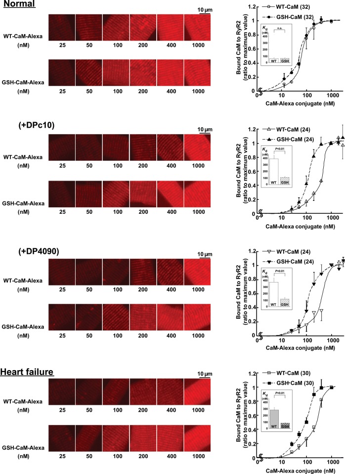
Figure 3.
The binding characteristics of exogenously introduced CaM in saponin-permeabilized cardiomyocytes. Delivery of various concentrations of CaM-Alexa or GSH-CaM-Alexa (left panel) and the summarized data (right panel) in normal cardiomyocytes and failing cardiomyocytes treated with RyR2 domain peptides (DPc10, 30 µmol/L, or DP4090–4123, 30 µmol/L). Either WT-CaM-Alexa or GSH-CaM-Alexa fluorescence was measured and expressed as the ratio to its maximum value. Each datum point per concentration represents mean ± SD of 8–10 cells from three to four hearts, and the sigmoid concentration-dependent relationships for CaM binding was fitted by an equation: y = aKnxn/(1 + Knxn), and the EC50 values were calculated as 1/K. (Inset) Comparison of EC50. Paired t-test was employed to determine the statistical significance of EC50. The numerical value in the parenthesis means the number of concentration-dependent relationships for CaM binding.

