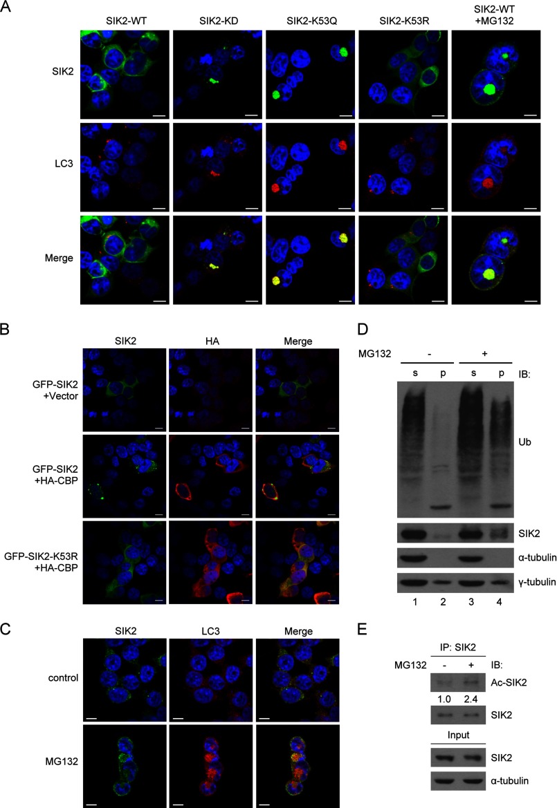
FIGURE 4.
Sequestration of acetylated-SIK2 to autophagosomes. A, HEK293T cells overexpressing the WT, KD, K53Q, or K53R variant of GFP-SIK2 were analyzed by immunofluorescence staining. Endogenous LC3 (red) was detected by anti-LC3B antibody. The rightmost panel shows GFP-SIK2-WT cells with MG132 (5 μm) treatment for 16 h. Scale bar, 10 μm. B, HEK293T cells were co-transfected with HA-CBP- (or the vector control) and GFP-SIK2-encoding plasmids, followed by immunofluorescence staining of the overexpressed HA-CBP (red) using anti-HA antibody. Scale bar, 10 μm. C-E, HEK293T cells were treated with or without MG132 (5 μm) for 16 h. They were then subjected to immunofluorescence staining (C), separated into soluble (s) and insoluble (p) fractions for protein expression analysis (D), or extracted for anti-SIK2 immunoprecipitation (E). The scale bar in C is equivalent to 10 μm. Western blots in D and E were done using the indicated antibodies. Relative levels of SIK2 acetylation were determined by normalizing the levels of acetylated SIK2 signals to those of total SIK2 protein in the respective samples, with the control sample being represented as 1.

