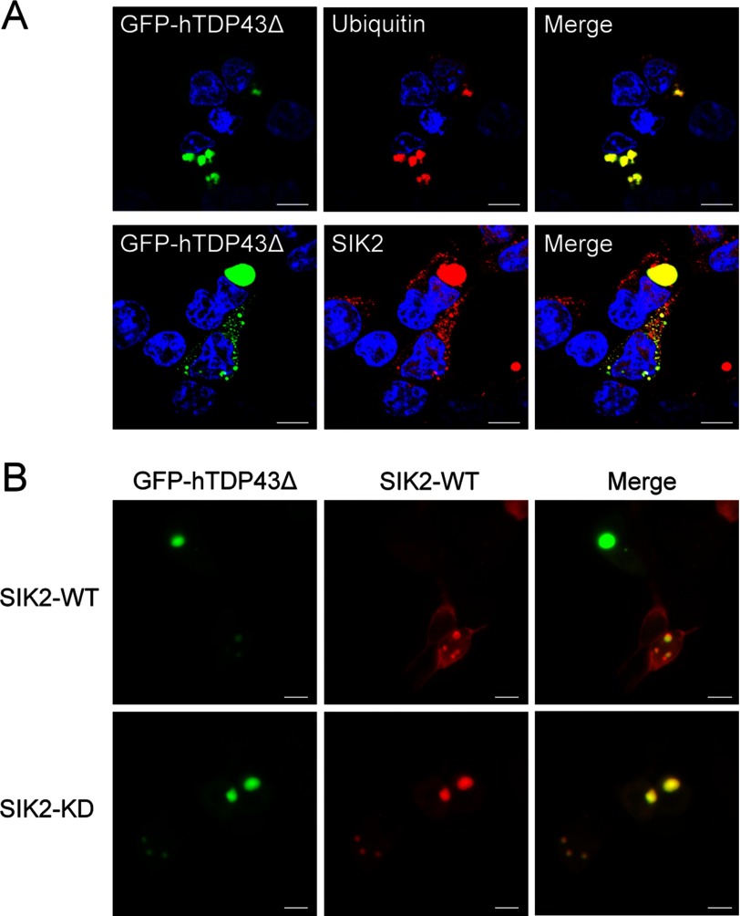
FIGURE 6.
SIK2 co-localizes with the TDP-43Δ inclusion bodies. A, HEK293T cells ectopically expressing GFP-hTDP43Δ were fixed and immunostained with anti-ubiquitin (upper) or anti-SIK2 (lower) antibodies. Scale bar, 10 μm. B, cells were co-transfected with GFP-hTDP43Δ- and FLAG-SIK2-encoding plasmids, followed by immunofluorescence staining using anti-FLAG antibody. Single-section images of confocal microscopy were acquired and shown (scale bar, 10 μm).

