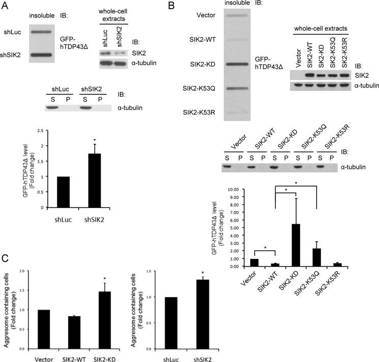
FIGURE 7.
SIK2 is required for the processing/removal of TDP-43Δ inclusion bodies. A and B, HEK293T cells were transfected with plasmids expressing luciferase- or SIK2-targeting shRNAs (A), or the WT, KD, K53Q, or K53R variants of SIK2 (B). GFP-hTDP43Δ was also co-expressed. Cell extracts were separated into soluble (S) and insoluble (P) fractions. Insoluble fractions were analyzed by slot-blot assay using anti-GFP antibody (top left panel). Soluble and insoluble fractions (middle) and whole cell extracts (top right panel) were probed with the indicated antibodies to show the expression levels of various proteins. Bar graph in the bottom panel shows the quantification of GFP-hTDP43Δ intensity on the slot-blot. Mean ± S.D. were calculated from 3 independent experiments. *, p < 0.05. C, HEK293T cells were transiently transfected plasmids for the co-expression of: GFP-hTDP43Δ with FLAG-SIK2 WT or KD (left panel) or GFP-hTDP43Δ with control shRNA or SIK2-targeting shRNA (right panel). Forty-eight h after transfection, cells were fixed and analyzed for the proportion of aggresome-containing cells. Quantitative data are mean ± S.D. of two independent experiments (*, p < 0.05).

