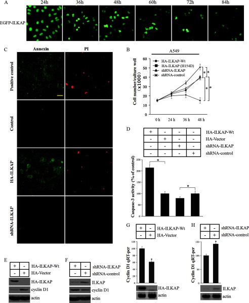
FIGURE 9.
ILKAP regulates cyclin D1 protein expression and induces apoptosis. A, HEK293 cells were transfected with the EGFP-ILKAP construct, and the expressed EGFP-ILKAP in the cells was visualized by UV microscopy at different times. B, A549 cells were transfected with HA-ILKAP, empty vector, shRNA-ILKAP or shRNA-control. The 3-[4,5-dimethylthiazol-2-yl]-2,5-diphenyltetrazolium bromide assay was performed to examine the cell proliferation rate 24, 36, and 48 h after the cells were transfected with each construct. C, the cells were assayed using the annexin V/propidium iodide (PI) apoptosis assay kit (Sigma-Aldrich) 48 h after the cells were transfected with each construct. D. The caspase-3 activity in the transfected cells was determined. E and F, immunoblots were performed to examine the cyclin D1 protein levels in the transfected cells. G and H, the RNA was extracted, and a quantitative RT-PCR assay was performed. * indicates a significant difference (p < 0.05) when compared with the control. Error bars indicate S.D.

