FIGURE 3.
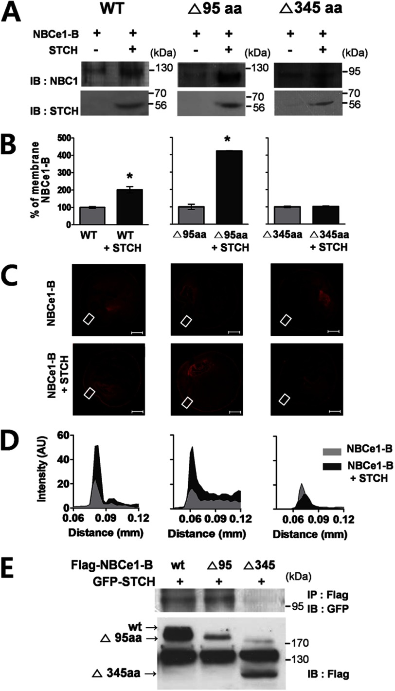
STCH binds to specific regions of NBCe1-B. A, Xenopus oocytes (n = 30 per transcript) injected with indicated cRNAs were extracted, and membrane fractions were subjected to Western blotting (IB) with indicated antibodies. Observations were repeated in at least two independent experiments for each construct: WT (n = 3), Δ95aa (n = 3), Δ345aa (n = 2). B, shown is densitometry quantification of bands illustrating wild type and mutant NBCe1 membrane expression in the presence of STCH as a percentage of control (without STCH) (*, p < 0.05). C, shown is immunolabeling of NBCe1-B wild type and deletion mutants in Xenopus oocytes transfected with NBCe1 alone (upper panels) or co-transfected with STCH (lower panels). Scale bars, 200 μm. Images are representative of two to three independent experiments for each transfection. D, shown is fluorescence intensity of membrane region corresponding to white boxed area in C. AU, absorbance units. E, HSG cells were transfected with FLAG-NBCe1-B, deletion mutants (Δ95aa, Δ345aa), and GFP-STCH plasmid. Cell lysates were subjected to immunoprecipitation (IP) with mouse anti-FLAG antibody, and immunoprecipitates were subjected to Western blotting with mouse anti-GFP antibody (n = 2).

