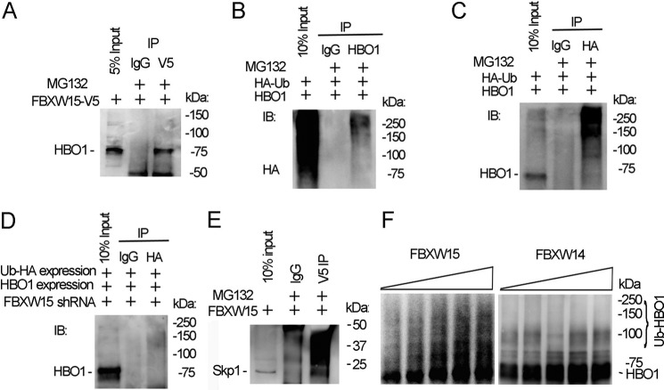
FIGURE 3.
Fbxw15 interacts with and ubiquitinates HBO1. A, pcDNA3.1/Fbxw15/V5 plasmid (4 μg) were transfected into MLE cells for 48 h. Cell lysates were subjected to V5 immunoprecipitation (IP). The immunoprecipitates were subjected to HBO1 immunoblotting (IB). B, pcDNA3.1/ubiquitin/HA plasmid (4 μg) and pcDNA3.1/HBO1/V5 plasmid (4 μg) were co-transfected into cells. Cell lysates (1 mg of protein) were subjected to HBO1 immunoprecipitation followed by HA immunoblotting. C, in reciprocal studies, cell lysates in B were immunoprecipitated with HA antibody, and the immunoprecipitates were analyzed by HBO1 immunoblotting. D, pcDNA3.1/ubiquitin/HA plasmids (4 μg) and pcDNA3.1/HBO1/V5 plasmids (4 μg) were co-transfected with Fbxw15 shRNA plasmids into cells for 48 h. Cell lysates (1 mg of protein) were subjected to HA immunoprecipitation followed by HBO1 immunoblotting. E, Fbxw15-V5 plasmid was overexpressed in cells, and lysates were subjected to V5 immunoprecipitation followed by Skp1 immunoblotting. F, HBO1 in vitro ubiquitination assays were conducted in the presence of Fbxw15 or a control, Fbxw14, and the full complement of ubiquitin reaction components. The samples were analyzed by HBO1 immunoblotting. The data are representative of three separate experiments.

