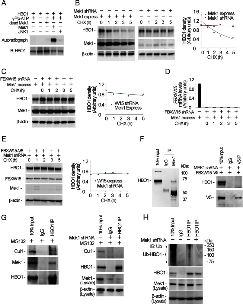
FIGURE 6.
Mek1 phosphorylates HBO1 and accelerates HBO1 degradation. A, HBO1 protein was subjected to in vitro Mek1 kinase phosphorylation assays. The lower panel shows HBO1 loading in each reaction as determined by immunoblotting (IB). B, Mek1 was overexpressed using Mek1 plasmid or knocked down by Mek1 shRNA in MLE cells for 48 h; the cells were treated with 20 μm of cycloheximide (CHX) for various time points. The cell lysates were analyzed by HBO1, Mek1, and β-actin immunoblotting. C, the cells were co-transfected with Mek1 plasmid and Fbxw15 shRNA plasmids for 48 h, and the cells were treated with 20 μm of CHX for various time points. The cell lysates were analyzed by HBO1, Mek1, and β-actin immunoblotting. D, Fbxw15 mRNA levels in the above Fbxw15 shRNA-treated cells were determined by quantitative RT-PCR. E, pcDNA3.1/Fbxw15/V5 plasmids (4 μg) were co-transfected with Mek1 shRNA plasmids into cells for 48 h. The cell lysates (1 mg of protein) were subjected to immunoblotting analysis as indicated. F, left panel, the cell lysates were subjected to Mek1 immunoprecipitation (IP) followed by HBO1 immunoblotting. Right panel, pcDNA3.1/Fbxw15/V5 plasmids (4 μg) were co-transfected with Mek1 shRNA plasmids into cells for 48 h, and V5 immunoprecipitation followed by HBO1 immunoblotting was performed. G, MLE cells were treated with MG132 (20 μm) for 1 h, and the cell lysates were subjected to HBO1 immunoprecipitation and followed by Cullin1, Mek1, and HBO1 immunoblotting (left panels). Mek1 was also knocked down with lentiviral shRNA constructs for 48 h, and the cell lysates were used for HBO1 immunoprecipitation followed by Cullin1 and HBO1 immunoblotting analysis. The cell lysates were also used for Mek1 and β-actin immunoblotting analysis (bottom panels). H, Mek1-depleted cellular immunoprecipitates were analyzed for ubiquitin and HBO1 immunoblotting after HBO1 immunoprecipitation. Bottom two panels, cell lysates were subjected to Mek1 and β-actin immunoblotting. In B, C, and E, results of densitometric analysis of immunoblots are represented graphically on the right. The data are representative of three separate experiments.

