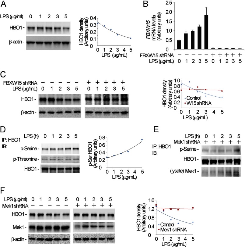
FIGURE 7.
LPS induces HBO1 degradation. A, MLE cells were treated with different concentrations of LPS for 18 h, and the cells were analyzed by HBO1 and β-actin immunoblotting (IB). B and C, scrambled RNA or Fbxw15 shRNA plasmids were transfected into cells for 48 h. The cells were treated with LPS at the indicated concentrations for 18 h. The cell lysates were subjected to HBO1 and β-actin immunoblotting analysis (C) and analysis of Fbxw15 mRNA levels by quantitative RT-PCR (B). D, the LPS-treated cells in A were immunoprecipitated (IP) with HBO1 antibody. The immunoprecipitates were probed with phosphoserine, phosphothreonine, and HBO1 antibodies. E, Mek1 knockdown cells were treated with LPS, and the cell lysates were immunoprecipitated with HBO1 antibody. The immunoprecipitates were probed with phosphoserine and HBO1 antibodies. The cell lysates were analyzed with Mek1 immunoblotting. F, cells were transfected with Mek1 shRNA plasmid or scrambled RNA for 48 h. The cells were treated with LPS at the indicated concentrations for 18 h. The cell lysates were subjected to HBO1, Mek1, and β-actin immunoblotting. In A, C, D, and F, the results of densitometric analysis of immunoblots are represented graphically on the right. The data are representative of three separate experiments.

