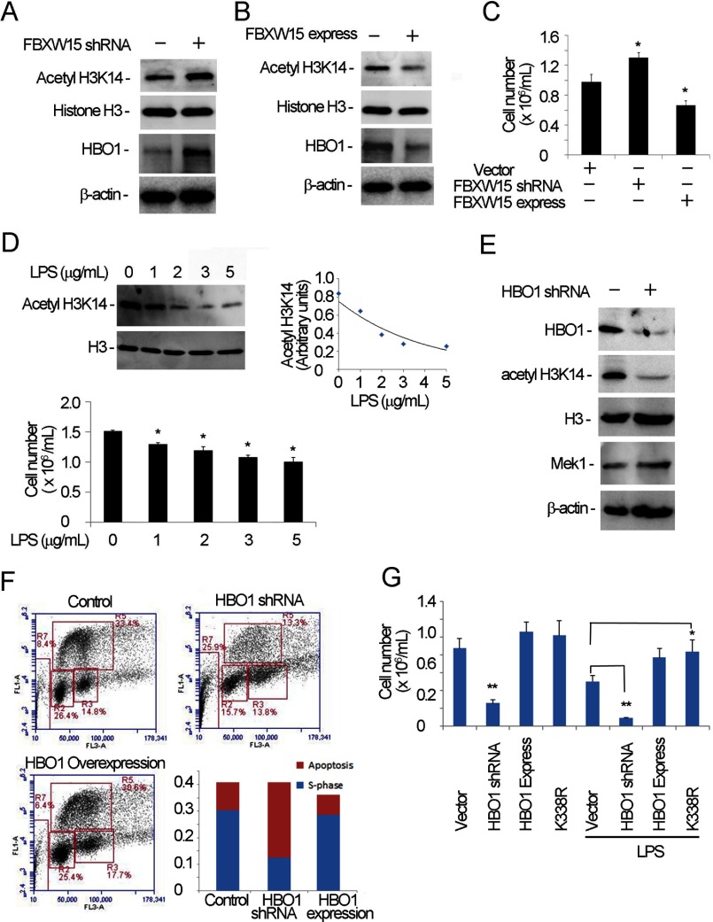
FIGURE 8.
Fbxw15 regulates histone H3K14 acetylation and cell proliferation. A and B, MLE cells were separately transfected with pcDNA3.1/Fbxw15/V5 (B) plasmids and Fbxw15 shRNA plasmids (A) for 48 h. The cell lysates were analyzed by H3K14 acetylation, histone H3, HBO1, and β-actin immunoblotting. C, treated cells from A and B were counted in cell proliferation assays. *, p < 0.05 versus control. D, cells were treated with various concentrations of LPS overnight, and the samples were subjected to anti-H3K14 acetylation immunoblot analysis (upper panels) and analyzed for cell proliferation (lower panel). The results of densitometric analysis of immunoblots are represented graphically on the right. E, HBO1 knockdown was conducted in MLE cells. The cell lysates were immunoblotted with the indicated antibodies. F, MLE cells were transfected with HBO1 plasmid or HBO1 was silenced in cells. The cells were subjected to FACS analysis. S phase and apoptotic cells were presented for different groups (lower right panel). G, cell proliferation was determined after HBO1 plasmid overexpression or silencing in cells. In HBO1 overexpressed cells, wild type HBO1 and K338R mutants were separately assayed. One set of the cells was treated with LPS (5 μg/ml) overnight. *, p < 0.05 versus vector control; **, p < 0.01 versus HBO1 shRNA. The data are representative of three separate experiments.

