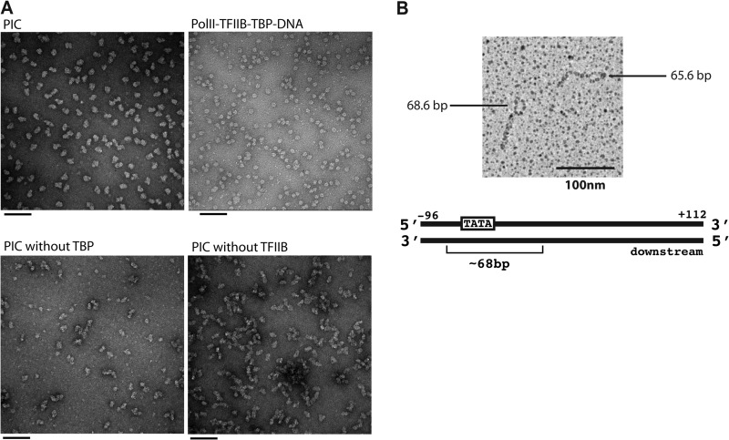
FIGURE 3.
Electron microscopy analysis of the PIC. A, representative images of negatively stained particles: the complete 31-polypeptide PIC with HIS4(−81/+19) (upper left), the pol II-TFIIB-TBP-DNA complex (upper right), the PIC assembled without TBP (lower left), and the PIC assembled without TFIIB (lower right). Scale bars = 100 nm. B, analysis of the PIC on the HIS4 promoter by psoralen cross-linking of DNA. The PIC with HIS4(−96/+112) was exposed to an interstrand DNA cross-linking agent. The DNA was then deproteinized and spread under fully denaturing conditions. The micrograph reveals an ∼68-bp bubble on one end, where proteins blocked access of the cross-linker to the DNA.

