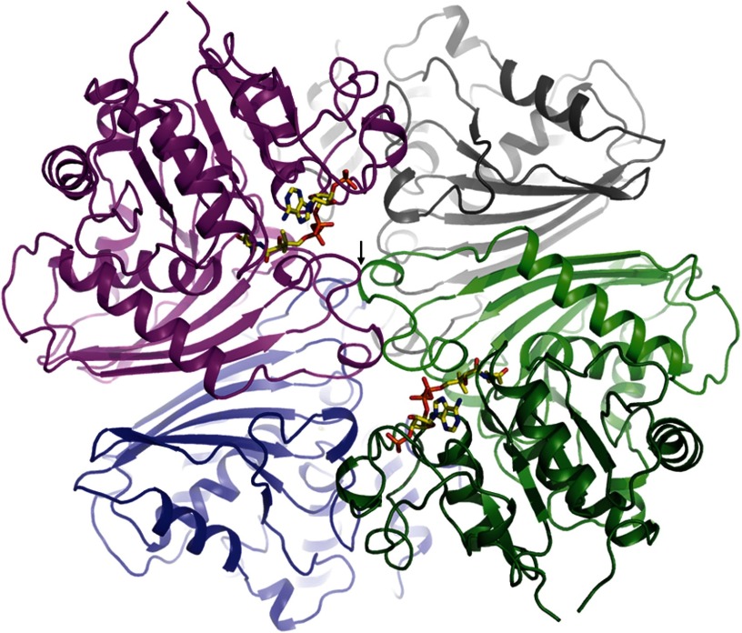
FIGURE 2.
Structure of MCR from S. todokaii. The MCR homotetramer is arranged as a dimer of dimers (colored in magenta/blue and gray/green). Each monomer is built up of a dinucleotide binding (dark green) and a dimerization domain (light green). The active site cleft lies between them and is occupied in the MCRCoA structure by CoA (drawn as a stick model). Two dimers form an extended interface that includes both cover loops (black arrow) being involved in substrate binding.

