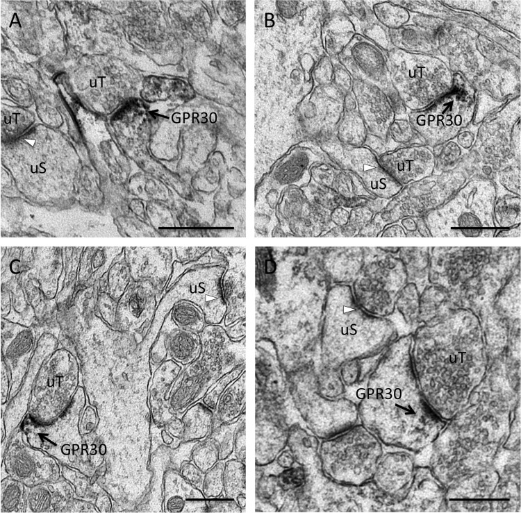
FIGURE 1.
GPR30 immunoreactivity in CA1 dendritic spine profiles. Shown are representative electron micrographs of GPR30 immunolabeling from the adult female rat CA1 hippocampus. Using pre-embedding electron microscopy, GPR30-labeled dendritic spines (GPR30) are identified by diffuse granular immuno-peroxidase reaction product and are indicated by black arrows. Unlabeled spines (uS) with unlabeled PSDs (white arrows) and unlabeled pre-synaptic terminals (uT) also are indicated. GPR30 IR is evident in multiple spine profile types, including thin spines (A), mushroom-shaped spines (B), cupped spines (C), and spines with perforated PSDs (D). GPR30-IR localized both at the PSD as well as in the peri-synaptic zone of the PSD. Panel A, at proestrus. Panels B–D, at diestrus. Bars, 500 nm.

