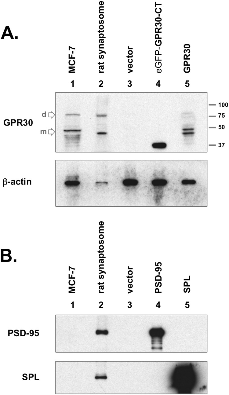
FIGURE 2.
GPR30 protein expression in the rat brain. A, upper blot, GPR30 was detected in crude rat brain synaptosome preparations (lane 2). COS-7 cells did not express GPR30, as GPR30 was not detected in empty vector-transfected COS-7 whole cell lysates (lane 3). GPR30 (C-terminal-specific) antibody recognizes the C-terminal domain of GPR30 (GPR30-CT), which was subcloned for chimeric expression with eGFP (lane 4). The rat full-length GPR30 protein are also detected when exogenously expressed in COS-7 cells (lane 5). MCF-7 whole cell lysate was used as a positive GPR30 antibody control (lane 1). The primary lower molecular weight GPR30 band is the monomeric protein state (m), and the upper secondary band at approximately twice the apparent molecular weight is likely the dimeric state (d) for the receptor. The apparent molecular weight ladder is shown on right. Lower blot, the same lysates were immunoblotted (IB) for actin protein content. Both blots are representative of five independent experiments. B, rat brain preparations contain synaptosomal proteins, including the PSD proteins PSD-95 and SPL. MCF-7 whole cell lysate (lane 1), rat brain synaptosome lysate (lane 2), or COS-7 whole cell lysates (lane 3, empty vector; lane 4, Myc-PSD95 transfected; lane 5, Myc-SPL transfected) were immunoblotted for PSD-95 protein (upper blot) or for SPL protein (lower blot). Blots are representative of three independent experiments.

